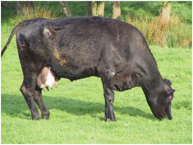
Gastrocnemius Muscle Rupture

- Rupture of the gastrocnemius muscle or tendon is relatively rare.
- It is most likely to be associated with deficiencies of calcium, phosphorus, and vitamin D.
- Prolonged recumbency, with resulting myositis and struggling to rise, occasionally precipitates rupture of these muscles.
CLINICAL SIGNS AND DIAGNOSIS

It presents as a markedly dropped (flexed) hock joint, sometimes reaching the ground, and a markedly flexed fetlock. It is particularly obvious in the standing animal. It is usually unilateral, and may become bilateral if the animal continues to struggle.
CheckOut The Video of Rupture of the Gastrocnemius Muscle
Treatment Of Rupture of the Gastrocnemius Muscle
Successful treatment is extremely unlikely in heavy adult animals. A leg cast or splint that maintains the hock in extension, supplying adequate vitamins and minerals, and proper nursing may be successful, but a long recovery period is required.

Tibial Nerve Paralysis

Etiology:
The tibial nerve is the caudal branch of the ischiatic nerve, which, in its proximal course, is well protected by the gluteal muscles. Distally, it progresses beneath the tendon of the gastrocnemius muscle and can be damaged when the tendon is traumatized. Tibial nerve injury is typically seen after prolonges second stage labour often caused by an oversized calf.
- Tibial paresis is commonly seen in bovine practice as a sequel to dystocia.
- The tibial nerve supplies the extensor muscles of the hock joint and the flexor muscles of the digits; affected cows are lame and have a dropped hock and knuckling of the fetlock.
- Complete functional recovery occurs not consistently after a conservative “wait and see” approach.
Clinical Findings:

- The hock joint is overflexed (dropped hock syndrome) and the fetlock is partially flexed.
- The gastrocnemius appears to be longer than normal and gives the impression that it or its tendon could be ruptured.
- Tibial nerve injury is typically seen after prolonges second stage labour often caused by an oversized calf.
- The fetlock tends to be buckled, but the animal can walk and bear weight, although its attempts to do so are awkward. Compared with that seen in peroneal nerve injury, the gait disturbance is mild, but the postural disturbance could be permanent.

CheckOut Video Of Tibial Nerve Paralysis
Treatment Abstract Of Tibial Nerve Paralysis
Eight dairy cows with tibial nerve paresis from different farms were presented for treatment. Seven cows had unilateral tibial nerve paresis and one cow, which was barely able to stand, had bilateral tibial paresis. The affected legs of the seven cows with unilateral paresis and the more severely affected leg of the remaining cow were stabilized using a cast made of synthetic resin. The claws and the skin of the affected limbs were cleaned and a thick layer of cotton was applied to pad the leg from the foot to the hock. A cast was then applied with the foot and metatarsus aligned in a straight line. The cast included the entire foot and extended to the hock. The cast was removed after 4 weeks. All of the eight cows could be kept in their normal environment. They were able to walk well with the cast and were only mildly lame. Feed intake and milk yield increased. After removal of the cast, seven of the eight cows walked normally, including the cow of which both legs had been affected. One cow was slightly lame with a dropped hock after cast removal but showed a normal gait 3 weeks later. In cows with tibial paresis, casting of the lower portion of the leg was a useful supportive treatment that resulted in restoration of normal gait. In seven of eight treated cows limb function was normal after 4 weeks, and in one cow after 7 weeks. This supportive therapeutic procedure is straightforward, minimizes aftercare and allows the cow to be kept in her normal environment.


![10 Question Banks Needed For ICAR AIEEA [Pdfs]](https://vetstudyhub.com/wp-content/uploads/2020/06/10-Question-Banks-Needed-For-ICAR-AIEEA-Pdfs-150x150.png)


Hi. The https://vets.petyaari.com site is great: it has a lot
of valuable information and is easy to find. I learned a lot from here, so I want to ask about a recommended book
as the best natural remedies: https://bit.ly/3cJNuy9
What do you think, it’s worth buying, is it
too cheap? Thanks and hugs!